One afternoon in 2000, Luo Qingming, then the associate dean of the School of Life Science and Technology at Huazhong University of Science and Technology(HUST), visited the office of Gordon Shepherd, a neuroscientist at Yale University in the United States. Shepherd was highly accomplished in fields such as neuroanatomy, neuronal networks, and neurocomputational science. Previously, Luo had an idea widely regarded as far-fetched, and on this occasion, he sought Shepherd’s opinion.
“ I want to pursue it. Do you think it holds any significance?”
“ It’s exceedingly meaningful.”
“The United States has both the conditions and the funding. Why haven’t you done it?”
“It’s too challenging. I might get fired before I even accomplish it.”
After talking with him, although Luo Qingming felt somewhat disheartened, he still decided to press on. Today, Luo Qingming has become a member of the Chinese Academy of Sciences and has transferred from HUST to Hainan University (HNU), where he serves as a professor of the School of Biomedical Engineering and the leader of the Brain Space Information Research Team. At HNU, he has finally turned the once seemingly “far-fetched” idea that had stumped Shepherd into a reality.
Luo Qingming has led his team to successfully create a mouse brain stereotaxic topographic atlas, enabling the generation of brain section images with isotropic 1-μm resolution at arbitrary angles. On July 2, the pioneering research findings were published in the journal Nature.
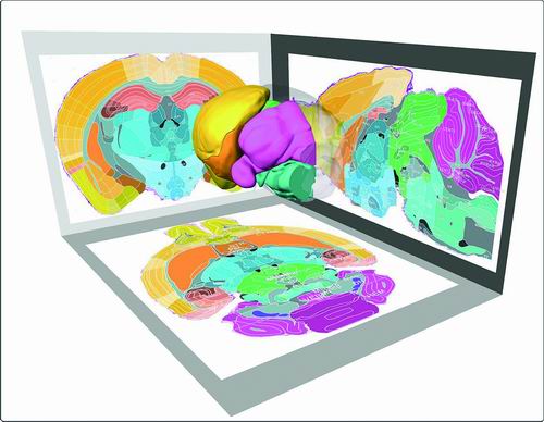
A mouse brain stereotaxic topographic atlas is mapped by Luo Qingming’s Brain Space Information Research Team. (Photo provided by the research group.)
The Brain’s Traffic: Why Mapping “Roads” Matters?
If we compare neurons in the brain to individual provinces, then neuronal conduction is akin to inter-provincial transportation. However, even with maps of Shandong and Hainan, the question of how to travel from Shandong to Hainan remains unanswered, as the routes connecting them are unclear. Brain scientists wonder not only where neurons are located in the brain and what they look like, but also how they achieve neuronal conduction. Yet, how can we “manage traffic”when the “routes” themselves are not even clear?
Previously, Luo Qingming’s team dedicated a full decade to developing a cutting-edge microscopic technology capable of capturing “3D full-body images” of biological tissues. This three-dimensional imaging system, named MOST (Micro-Optical Sectioning Tomography), excels at visualizing the intricate structures of organs such as brain, even revealing the connectivity patterns of neurons. In essence, they had acquired a tool to unravel the “uncharted territories” of the brain.
In 2010, their corresponding research paper was published in the journal Science, marking the first Science article in instrumental development research not only for HUST but also for China as a whole.
At that time, some advised Luo Qingming to cease to do it. After all, it seemed somewhat fanciful for a physicist to attempt mapping a three-dimensional atlas of a mouse brain. Moreover, since the early 21st century, scientists worldwide had been obsessively pursuing three-dimensional brain atlases, pushing slice thickness from 40μm micrometers down to 25μm then to 10μm. Even the U.S. BRAIN Initiative had once assembled a team of researchers to strive for ultimate precision, only to end in failure.
Nevertheless, Luo Qingming remained undeterred, choosing instead to challenge the next summit—developing a stereotactic atlas of the mouse brain with single-cell resolution. With approximately 90% of its genes homologous to humans, the mouse shares striking physiological and pathological similarities with our species. Given that the human brain is 1,000 times more complex than its murine counterpart, no major scientific project currently directly supports human brain atlas development. However, MOST technology, the high-resolution three-dimensional imaging system, could capture images 100 times clearer than traditional methods, rendering conventional mouse brain atlases obsolete and making the creation of a new three-dimensional brain atlas feasible.
Luo Qingming set his sights on achieving 1-μm slice thickness and arbitrary-angle sectioning. How challenging was this goal?
Li Yunqing, President of the Chinese Society for Anatomical Sciences, explained to China Science Daily that 1μm is equivalent to 1/50th the diameter of a human hair. Yet this seemingly minuscule 1-μm slice thickness enables visualization of neuronal interiors— a feat unattainable with previous technologies, which maxed out at 10-μm slice thickness and 100-μm axial resolution, insufficient for distinguishing individual cells. The necessity of arbitrary-angle sectioning is most vividly demonstrated in applications like B-ultrasound examinations,as the ultrasound probe rarely contacts the human body at exactly 90 degrees—it may be at 30 degrees, 135 degrees, or indeed any other angle. Only by enabling cross-sectional imaging at arbitrary angles can more accurate diagnostics be achieved.
A Decade-Long Odyssey in Brain Mapping
With a background in physics, Luo Qingming never presumed to call himself a neuroscientist. Yet upon completing this groundbreaking research, observers noted that his entire team had effectively mastered neuroanatomy.
In 2010, core team member Feng Zhao was completing his bioinformatics PhD at HUST, deeply immersed in genetic research. His introduction to Luo Qingming’s project came as a surprise: “I never anticipated becoming a specialist in neural cartography,” he reflected.
Traditional brain atlases present blurry two-dimensional section information—like a flat paper map—unable to fully showcase the three-dimensional form, size, and position of neural cells or the connections between neurons. The team’s goal was to make this map three-dimensional and dynamic.
“The first thing we did was manually sketch brain region diagrams,” said Feng Zhao. The atlas he initially used was a 100-page booklet, then regarded as the “gold standard”: 25μm-thick sections, 100-pixel axial resolution, and only coronal and sagittal planes (effectively just X and Y axes) with coarse image quality. These constraints collectively rendered 3D modeling from this atlas nearly impossible.
To achieve 1μm resolution and arbitrary-angle slicing, the researchers needed to establish a comprehensive three-axis coordinate system. Years of painstaking work yielded over 10,000 meticulously hand-drawn brain region maps. Their expertise became so refined that “they could instantly identify any brain region and position from a randomly chosen sketch.” With the integration of machine learning, their mapping process accelerated dramatically. Otherwise, “manually sketching everything from scratch would have taken 20 years.”
A decade later, a transparent “crystal brain” of a mouse appeared on their computer screen—stitched together from 14,000 coronal slices, 11,400 sagittal slices, and 9,000 transverse slices, forming a cytoarchitecture image of the entire mouse brain. After staining, 916 brain regions became visible, including 236 newly discovered subregions that revealed previously unknown neural connectivity networks.
When asked why the research took so long, Luo Qingming often replied, “The only honest answer is that we simply weren't clever enough to find a faster way.” Now, having trained his students into experts, no one questions a physicist’s capability to map a mouse brain.
A Legacy of Precision
While the research required a decade to complete, Luo Qingming believes its impact will be far-reaching, particularly in fields like brain-computer interfaces (BCIs) and artificial intelligence (AI).
The recent success of Elon Musk’s neural implant technology, which restored speech capabilities of mute patients, highlights the critical importance of precise neural mapping. “For invasive implants, electrode placement accuracy is paramount,” Luo emphasized. “Our atlas provides the micron-level spatial resolution needed for truly targeted interventions.” He added that while the fundamental principles underlying the human brain atlas and the mouse brain atlas are aligned, advancing human brain mapping requires support from large-scale national scientific initiatives.
Team member Gong Hui of Wuhan National Laboratory for Optoelectronics, HUST, explained their approach: “Mouse models provide an essential experimental platform for understanding neurological disorders.” Their atlas has already yielded significant discoveries, including memory-encoding circuitry conserved in both rodents and humans, and precisely mapped substantia nigra dopaminergic neuron clusters relevant to Parkinson’s disease—enabling targeted optogenetic and pharmacological interventions.
While the team’s brain atlas sets a new “gold standard” for the field, they chose open-source accessibility over proprietary control. A visualization and sharing platform in Suzhou now offers cloud computing and public data downloads. Li Yunqing elaborated: “This means junior surgeons can locate brainstem hemorrhages by using the atlas, and neurosurgeons can simulate procedures with unprecedented accuracy before operations.”
The scientific community has welcomed this achievement enthusiastically. Prof. Xue Tian from the University of Science and Technology of China (USTC) immediately messaged the team: “The field has relied on the Paxinos & Franklin atlas for decades. Now we have a new precision benchmark developed in China.”
Javier DeFelipe, a reviewer from Spain’s Cajal Institute (CSIC), offered this assessment: “This work provides a powerful neuroinformatics tool for brain research at the single-cell level, marking a monumental achievement.”
Translated by Lin junjun, Ye Kechen
Proofread by Feng Jun, Wang kexian